Skip to content
For Physicians For Patients
Stratus Medical
Clinical Evidence

Clinical References, Evidence & Validation.

Peer-reviewed studies, technical validations, and case reports on the NIMBUS Multitined Expandable Electrode across anatomies.

1
Seminal Scientific Article

Ideal lesion characteristics, device description, & lesion size of NIMBUS MEE

View Article
2
NIMBUS MEE for Lumbar RFA

Clinical advantages v. conventional technique, reducing procedure time by 50%

View Article
3
Other References

Comparison of Lesion Volumes & Shapes Produced with Cooled, Protruding, & Standard RFA

View Article
4
Monopolar Lesion Size & Shape

Characterizes the size & shape of the NIMBUS MEE lesion as 467 mm3

View Article
5
NIMBUS MEE for Lumbar RFA

Perpendicular placement of NIMBUS MEE for lumbar MRI validated to be 601 mm3

View Article
6
NIMBUS MEE for SIJ RFA

Bench science & post-procedure MRI validation of size & shape of strip lesion for SIJ RFA

View Article
7
NIMBUS MEE for SIJ RFA

Case report with MRI validating the size & shape of strip lesion for SIJ neurotomy

View Article
8
NIMBUS MEE for SIJ RFA

12 month outcomes in 182 patients utilizing NIMBUS MEE for SIJ RFA

View Article
9
NIMBUS MEE for SIJ RFA

SIJ innervation from S1–S3 LBs varies and benefits from a large-volume lesion

View Article
10
NIMBUS MEE for Lumbar RFA

Coaxial (94 p.) to Multifidus Sparing (401 p.) Lumbar RFA with >56% of patients reporting positive outcome at 1 year

View Article
11
NIMBUS MEE for SIJ RFA

Significant pain reduction sustained up to 9 months with ultrasound-guided RFA using NIMBUS MEE

View Article
12
History of RFA Neurotomy

Advancements in RF technology, including NIMBUS Multitined Expandable Electrode

View Article
13
Other References

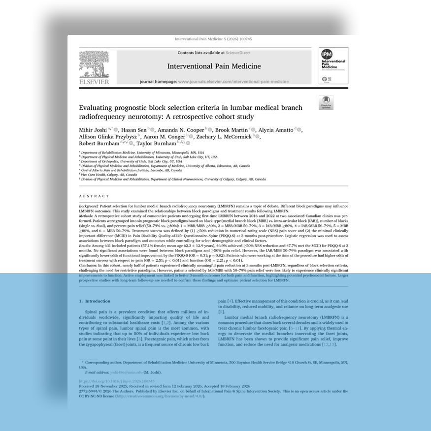
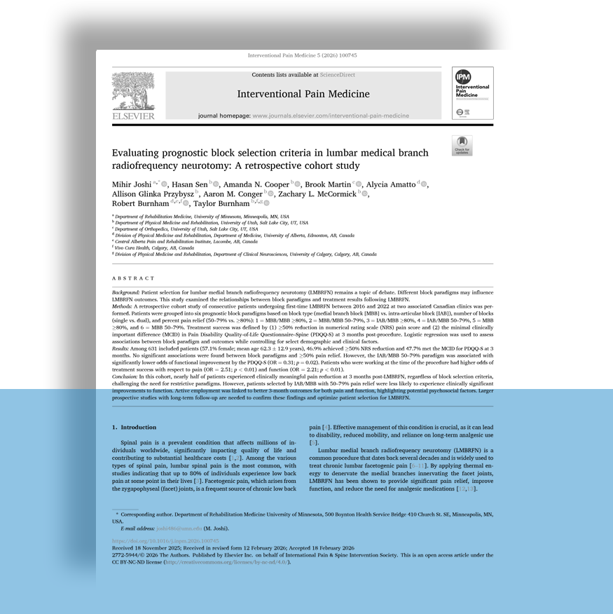
Consensus Practice Guidelines on Interventions for Lumbar Facet Joint Pain from a Multispecialty, International Working Group

View Article
14
ASPN Updated Guidelines for RFA

Supports either multiple lesions or large volume lesion

View Article
15
NIMBUS MEE for SIJ RFA

12 month outcomes in 182 patients utilizing NIMBUS MEE for SIJ RFA

View Article
16
Large Volume Lesion RFA for Genicular

Statistically significant decrease in pain intensity were observed at up to 24 months

View Article
17
Angle of Insertion for Lumbar RFA

Parallel Placement is Technically Challenging - L4 was 31.04º ± 1.83º & L5 was 40.74º ± 1.86º

View Article
18
NIMBUS MEE v Standard Cannula

Muscle-Bone Interface in Ex-Vivo Model Comparison

View Article
19
Other References

Advanced Interventional Procedures for Knee Osteoarthritis

View Article
20
RFA for Infrapatellar Knee Pain

Image-guided technique for targeting the infrapatellar branch of the saphenous nerve

View Article
21
Diagnostic & RFA Techniques for SIJ

Review of Sacral anatomy, diagnosis, & RFA techniques

View Article
22
NIMBUS MEE for Genicular RFA

At a mean follow-up of 9.0 ± 1.5 months, 50.0% reported ≥50% NRS reduction and 56.3% reported PGIC scores ≥6

View Article
23
RFA for Greater Trochanteric Hip Pain

≥50% NPRS reduction reported by 55.6% of participants at a mean follow-up of 13.1 months

View Article
24
RFA for Genicular Pain

Larger lesions increase the likelihood of capturing the targeted structure

View Article

Bring NIMBUS into your practice.

Request a demo or in-service, and our clinical team will walk your staff through it.

Get in touch
Stratus Medical
32303 FM-2978 · Magnolia, TX 77354
info@stratusmedical.com · 346-703-0642
© 2026 Stratus Medical. NIMBUS®, Vesta®, Stratus® Medical, and Advancing RF Technology for the Treatment of Pain® are trademarks of Stratus Medical. All illustrations are for demonstration purposes only.  ECO-10198 MKT-026 Rev. B